Introduction
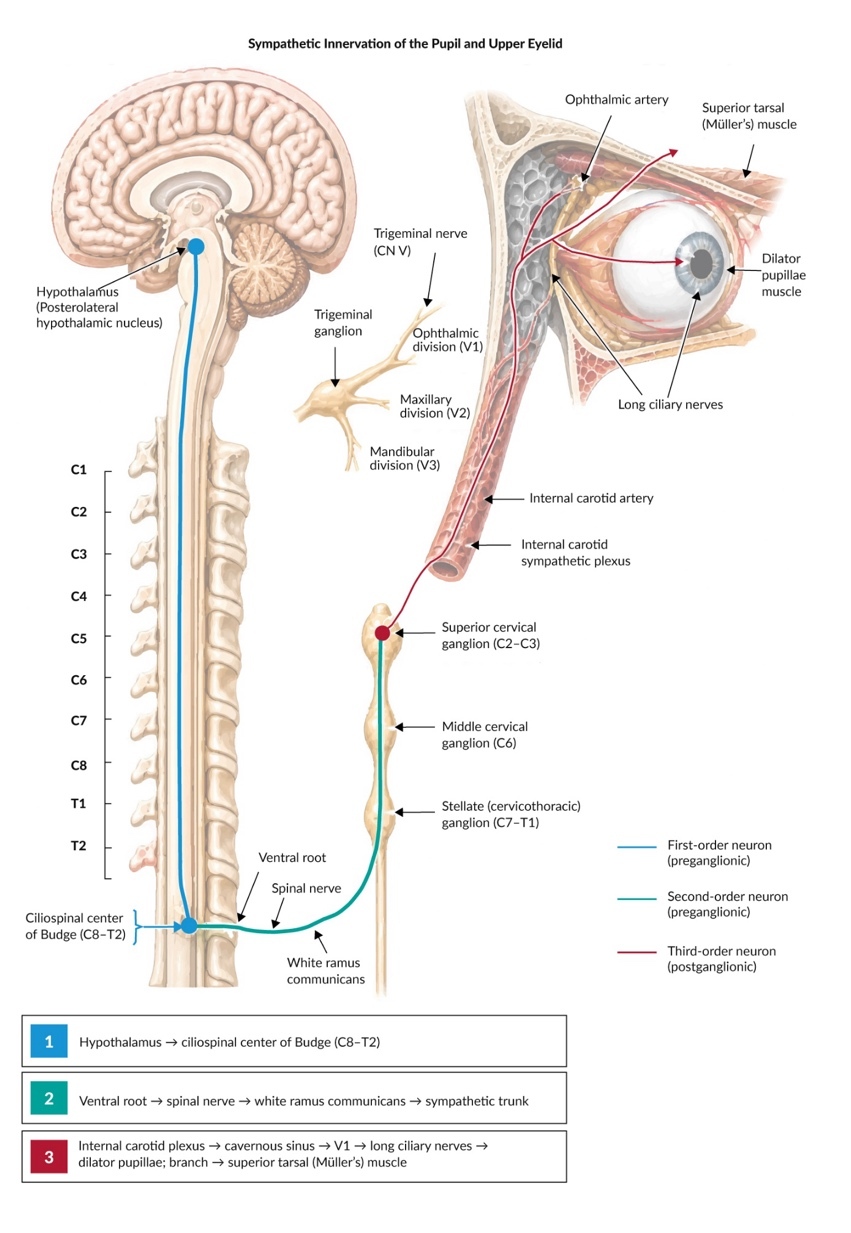
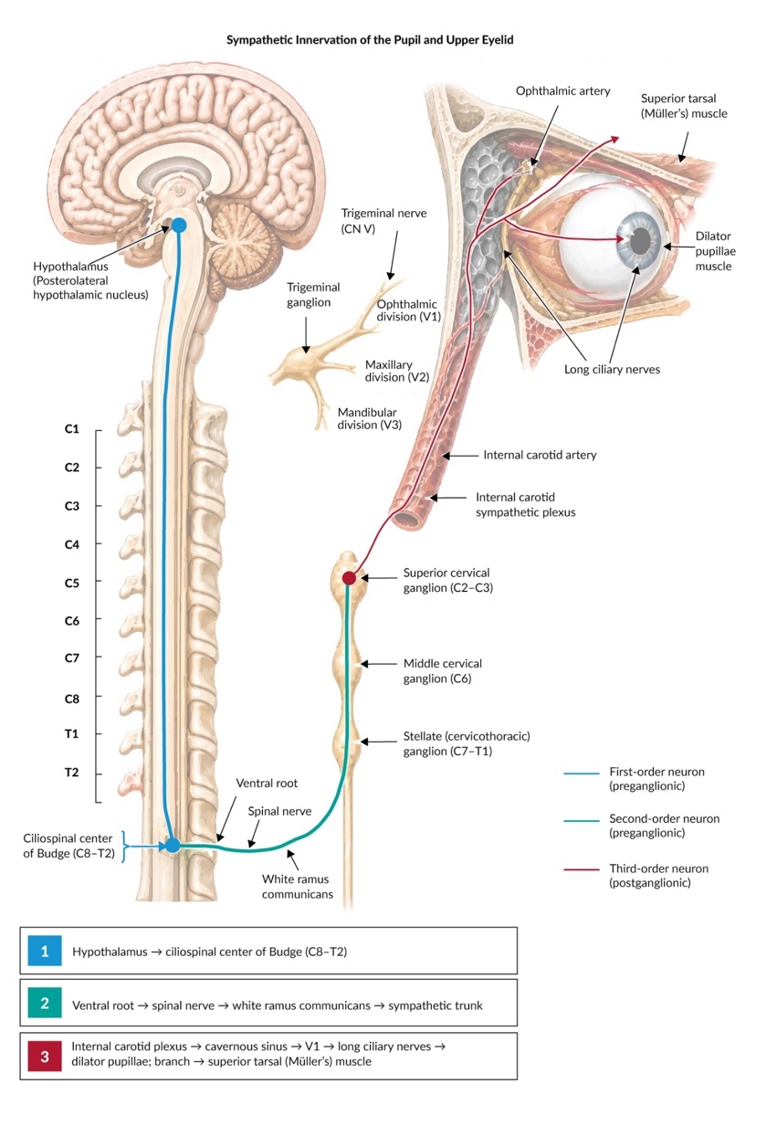
Horner syndrome results from interruption of the oculosympathetic pathway, which carries sympathetic innervation to the eye and periocular structures.1 Disruption of this pathway produces the classic triad of ptosis, miosis, and anhidrosis, although the presence and extent of anhidrosis are highly variable and depend on the anatomical level of the lesion.2 In postganglionic lesions, clinically evident anhidrosis is typically absent or restricted to a small area of the forehead, as most sudomotor fibers branch along the external carotid artery before this level.3,4 Anatomically, the pathway extends from the hypothalamus to the ciliospinal center, the superior cervical ganglion, and finally the eye and facial sweat glands (Figure 1).1,5–8
The clinical presentation varies depending on lesion location, with hallmark findings including anisocoria that becomes more pronounced in dim lighting as the affected pupil fails to dilate appropriately.9 Horner syndrome can be congenital or acquired, with acquired cases often resulting from trauma, vascular pathology, neoplasms, or iatrogenic injury.10,11 Internal carotid artery dissection is an important cause of acute Horner syndrome in adults and warrants urgent neurovascular assessment. While acute traumatic cases are well-documented in neurosurgical12 and ophthalmological literature,13 long-standing post-traumatic Horner syndrome is less frequently described, particularly from a primary care perspective.14
We present a case of post-traumatic Horner syndrome with a twelve-year interval between initial injury and diagnosis, highlighting how primary care recognition of specific upper and lower eyelid signs initiated targeted diagnostic work-up. In this patient, surgical correction of Horner-associated ptosis provided functional, cosmetic, and patient-reported benefit after a prolonged diagnostic interval. Subtle presentations may first be encountered in primary care, where recognition is important for appropriate referral and intervention.
Case Presentation
A 43-year-old male presented to our family medicine clinic for routine health assessment in 2021. During examination, we observed left-sided upper eyelid ptosis that the patient had not mentioned previously. Upon direct questioning about this finding, he reported persistent left-sided pupillary asymmetry following a penetrating workplace injury in 2009, when a metallic foreign body became embedded in the supraclavicular region, necessitating arterial surgery. He developed left-sided ptosis and miosis after the injury and subsequent surgery but was never evaluated specifically for these symptoms, as management focused entirely on the vascular injury. The patient reported no associated pain, diplopia, or other neurological symptoms.
Patient information/personal history
His medical history was otherwise unremarkable, with no chronic conditions or medications. He had relocated to Switzerland in 2019. During the consultation, he reported that the facial asymmetry affected his self-confidence, particularly in professional settings.
Clinical examination
Physical examination revealed a typical, moderate ptosis of the left upper eyelid, with a Margin Reflex Distance 1 (MRD1) of 2 mm on the left compared to 4 mm on the right, representing a 2 mm upper lid asymmetry. Levator function was normal bilaterally (14 mm). Notably, a mild elevation of the left lower eyelid (“upside-down ptosis”) was also present. This reliable indicator of sympathetic denervation narrows the palpebral fissure from below and frequently creates a false impression of true enophthalmos. Preoperatively, a slightly increased compensatory recruitment of the frontalis muscle was noted via discrete elevation of the left eyebrow.
Pupillary examination under ambient illumination demonstrated a discrete miosis of the left pupil (2.5 mm compared to 4 mm on the right). In dim lighting, the resulting anisocoria became more pronounced (2.0 mm left vs 4.5 mm right) due to a characteristic dilation lag of the affected pupil. Both direct and consensual pupillary light reflexes were intact and brisk bilaterally, and a swinging-flashlight test revealed no relative afferent pupillary defect (RAPD). Facial sweating was reported as symmetrically normal by the patient, which is common as anhidrosis is frequently subclinical depending on the exact composition of injured fibers. The remainder of the cranial nerve examination was unremarkable, with no evidence of other neurological deficits.
The chronicity of symptoms, clear temporal relationship to trauma, and classic presentation strongly suggested a post-traumatic Horner syndrome. Given the approximately 12-year interval between the injury and diagnostic evaluation, coupled with the patient’s functional concerns, we initiated a systematic evaluation to confirm the diagnosis, exclude relevant structural or vascular causes, and explore treatment options.
Diagnostic assessment
Based on clinical suspicion of Horner syndrome, we referred the patient for neurological consultation at the University Hospital Basel. Neurological and neuro-ophthalmological examination confirmed our findings of moderate left ptosis and miosis without additional neurological deficits. Pharmacologic testing with 0.5% apraclonidine demonstrated reversal of anisocoria, with significant dilation of the affected pupil and minimal effect on the unaffected side, confirming denervation hypersensitivity characteristic of Horner syndrome.15,16
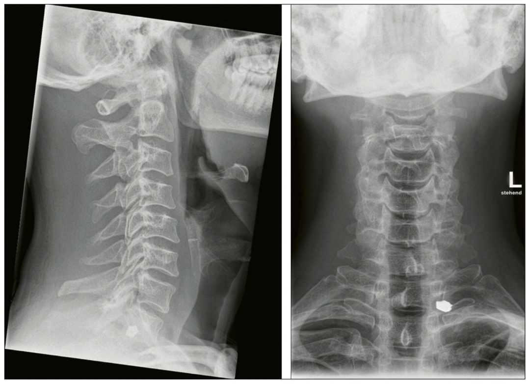
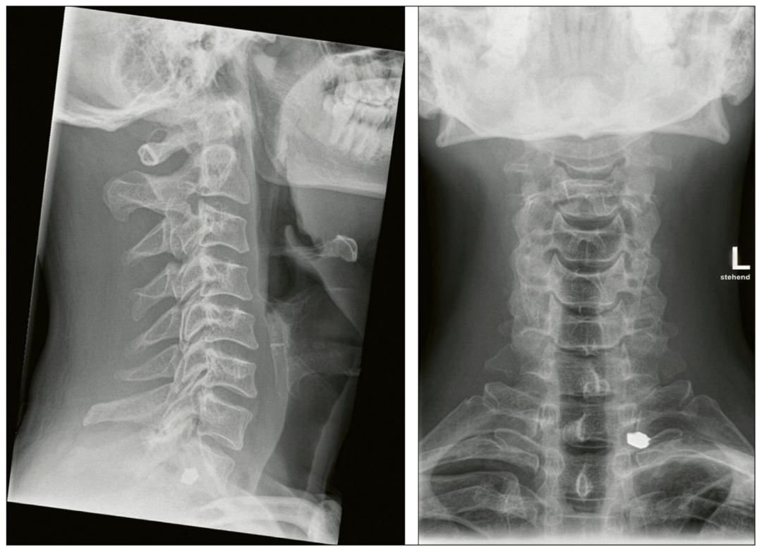
To evaluate potential structural causes and support anatomical localization of the lesion, contrast-enhanced magnetic resonance imaging (MRI) and magnetic resonance angiography (MRA) of the brain, cervical spine, and upper thorax were performed. Given the remote history of penetrating supraclavicular trauma and subsequent vascular surgery, imaging was obtained to characterize the regional anatomy and assess for residual structural abnormalities or delayed vascular sequelae along the sympathetic pathway before surgical planning. Before MRI, historical surgical records were reviewed to verify that the retained supraclavicular metallic material, visible on the radiographs (Figure 2), was composed of non-ferromagnetic alloy.
The neuroimaging revealed no acute pathology, no mass lesion, active inflammation, or vascular abnormality (such as an aneurysm or late dissection). However, it demonstrated clear evidence of residual post-traumatic soft tissue changes adjacent to the left sympathetic chain at the level of the supraclavicular region, consistent with the patient’s history. Cervical spine radiographs further documented the retained left-sided metallic foreign body projecting at the T1/T2 level (Figure 2).
The combination of clinical presentation, pharmacologic confirmation, and neuroimaging findings supported the diagnosis of post-traumatic Horner syndrome, most compatible with involvement of the third-order sympathetic pathway.17 The benign, stable nature of the findings, coupled with the long-standing chronicity, indicated no requirement for acute neurosurgical intervention.
Therapeutic intervention
Following confirmation of a stable, chronic post-traumatic Horner syndrome, management focused entirely on the surgical correction of the symptomatic ptosis, which impacted the patient’s quality of life and professional interactions. No intervention was indicated for the asymptomatic miosis. Through interdisciplinary collaboration between neurology, ophthalmology, and primary care, the patient was evaluated for surgical ptosis correction. Before surgery, a phenylephrine test (2.5%) was performed, which yielded only a partial response of upper eyelid elevation.
The patient was referred to the ELZA Institute for ophthalmological intervention. Given the specific presentation of sympathetic denervation affecting Müller’s muscle with preserved levator function, a tarsoconjunctival müllerectomy was planned.18 To achieve complete vertical symmetry and compensate for a suspected mild secondary aponeurotic component due to the 12-year chronicity of the condition, a concurrent limited levator shortening was intraoperatively performed by the oculoplastic surgeon. This tailored approach targeted the Horner-associated ptosis components directly.
Follow-up and outcomes
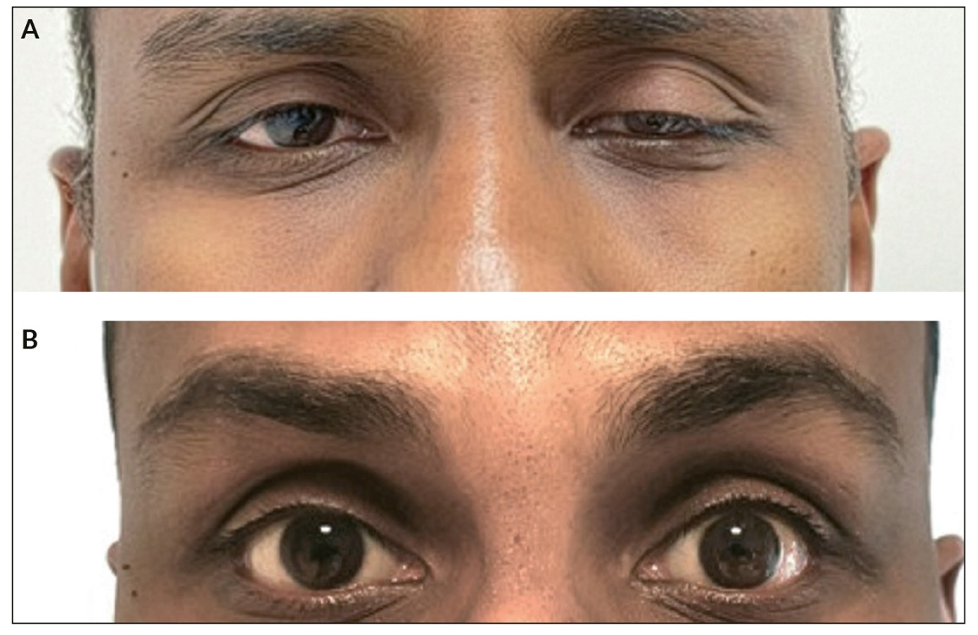
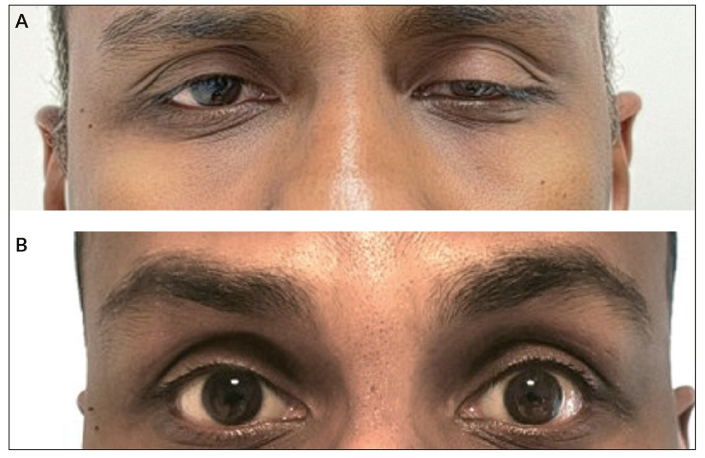
Postoperative follow-up evaluations at one, three, and six months demonstrated favorable surgical outcomes. Postoperative MRD1 was 4.5 mm on the left and 4.0 mm on the right, leaving a 0.5 mm difference (Figure 3A and B). Normal ocular surface integrity and symmetrical lid closure were maintained, with no complications such as lagophthalmos, dry eye syndrome, or corneal exposure.19
Beyond the objective improvements, the patient reported significant enhancement of self-confidence and satisfaction with his appearance. Using a visual analog scale to assess subjective improvement (0–10), the patient rated his satisfaction as 9/10 compared to 3/10 preoperatively. He specifically noted improved comfort in professional and social interactions, highlighting the psychological impact of successful intervention for this visible sequela of Horner syndrome.
Discussion
This case provides several important insights into the recognition and management of subtle neuro-ophthalmological findings in primary care. In particular, it demonstrates that long-standing Horner-associated ptosis can be treated surgically, as illustrated by the outcome in this patient.
Traumatic Horner syndrome represents approximately 4–13% of all Horner syndrome cases, with most literature focusing on acute presentations associated with carotid dissection or direct sympathetic chain injury.1,11 Our case bears similarity to the report by Guillén-Paredes et al. of penetrating cervical trauma resulting in Horner syndrome without vascular injury, although their case was diagnosed acutely and involved additional cranial nerve findings.20 The extended interval between injury and diagnosis in our patient offers a practical perspective on the delayed recognition and subsequent management of long-standing post-traumatic sympathetic disruption.
The primary care approach to neuro-ophthalmological conditions requires systematic clinical reasoning and appropriate use of diagnostic resources.21 In this case, recognition of upper and lower eyelid signs during routine examination triggered targeted questioning that revealed the historical context necessary for clinical suspicion.22 This emphasizes the value of thorough physical examination in primary care, particularly for detecting signs that patients may not identify as significant.23
In the primary care setting, a key clinical maneuver to differentiate Horner syndrome from other causes of anisocoria is the assessment of “dilation lag”. Because the pupillary dilator muscle is paretic, the affected pupil dilates more slowly when ambient light is removed, making the anisocoria most pronounced within the first 4 to 5 seconds of darkness.9 Emerging technologies, such as automated video pupillography devices tailored for primary care (e.g., neos by machineMD), now allow for objective, automated measurements of pupil size and dilation kinetics. In the future, the integration of such tools could enable definitive pupillary screening directly within the primary care setting, potentially reducing the need for specialist referrals in stable or chronic cases.
Pharmacologic testing remains a cornerstone of non-invasive diagnosis. Apraclonidine (0.5% or 1.0%) relies on denervation hypersensitivity; it not only reverses pupillary anisocoria by dilating the miotic pupil but typically corrects or overcorrects the Horner-associated ptosis due to its alpha-adrenergic effect on Müller’s muscle.16 While our patient was referred to a tertiary center, the apraclonidine test is a straightforward diagnostic tool that can be safely performed in secondary care or appropriately equipped primary care clinics, bypassing the need for specialized neurological consultation in clear, non-acute clinical scenarios.
This case helps define a clear referral paradigm for primary care physicians encountering suspected Horner syndrome. The management pathway depends strictly on acuity and etiology:
-
Urgent Neurovascular/Neurological Assessment: Any acute, painful, or new-onset Horner syndrome – even without a history of trauma – requires immediate emergency referral for neuroimaging (CTA/MRA) to rule out life-threatening internal carotid artery dissection or neoplastic compression.
-
Oculoplastic/Ophthalmological Referral: Conversely, in patients with a long-standing, completely stable presentation and a clearly established benign etiology (such as remote trauma or iatrogenic injury), extensive neurological work-up holds limited value. These patients can be referred directly to an oculoplastic surgeon for symptomatic ptosis management, streamlining care and reducing healthcare costs.
Several limitations must be acknowledged in interpreting this case. As a single case report, generalizability is inherently limited. Patient-reported satisfaction was assessed using a simple visual analog scale rather than a validated quality-of-life instrument. Furthermore, while our extensive work-up included advanced neuroimaging, a more tailored and cost-effective diagnostic pathway could be considered in similar clinical scenarios. Specifically, patients presenting with a long-standing, stable Horner syndrome and a clear, documented traumatic or surgical etiology can potentially be referred directly to an oculoplastic surgeon or neuro-ophthalmologist, bypassing extensive neurological testing and MRI. Conversely, acute presentations or cases without an obvious cause represent a potential neuro-vascular emergency (such as carotid artery dissection) that warrants immediate neurological evaluation and urgent imaging. In addition, objective photographic documentation of the positive apraclonidine test was not captured preoperatively. Nevertheless, the classic presentation, clear temporal relationship to trauma, and favorable outcome after ptosis correction support the diagnosis and management approach.
Conclusion
This case illustrates that primary care recognition of specific upper and lower eyelid signs can initiate appropriate diagnostic work-up and specialist referral, even when symptoms have been present for many years. While the underlying sympathetic disruption itself is not reversed, delayed surgical correction of Horner-associated ptosis was associated with functional, cosmetic, and patient-reported improvement in this patient.
Ethical considerations: Consent and privacy
This case report was prepared in accordance with the principles of the Declaration of Helsinki and with ethical standards for medical publication and patient confidentiality. The patient provided written informed consent for publication of this case report and the accompanying clinical images, including the radiographs and clinical photographs shown in Figures 2 and 3. The patient was informed that, despite anonymization, complete anonymity cannot be guaranteed because clinical images are included. All patient information was anonymized as far as possible.
Availability of data and materials
All relevant patient data supporting this case report are included in anonymized form within the published article.
Conflict of interest
The authors declare that this case report was prepared in the absence of any commercial or financial relationships that could be construed as a potential conflict of interest.
Funding
All authors have declared that no financial support was received from any organization for the submitted work.
Author contributions
All authors contributed to and approved the final manuscript.