Loading [Contrib]/a11y/accessibility-menu.js
1.
Fetian IR, Fitian ID, Villiger L, Darwish M. Response to Nigella sativa in Patients with Confirmed and Suspected COVID-19. hb TIMES Schw Aerztej. 2020;1(1):66-75. doi:10.36000/hbT.2021.01.001
Download all (7)
  • Figure 1. CRP measurement: Proven and exclusive immunoturbidimetry technology for a rapid and accurate diagnosis.
  • Table 1. Active components of Nigella sativa.
  • Figure 2. Number of people testing positive by gender.
  • Figure 3. Number of people who tested positive for COVID-19.
  • Figure 4. Effect of NS Treatment on CRP.
  • Figure 5. Endotheliitis. Adapted from Varga et al. 2020.
  • Figure 6. Possible mechanisms for the actions of NS against COVID-19.

Abstract

Coronavirus Disease 2019 (COVID-19) has been widely spreading all over the world since the beginning of 2020. World Health Organization, on January 30th, 2020, declared the spread of COVID-19 to be a “public health emergency of international concern” (PHEIC). As of January 8th, 2021, 89,107,341 COVID-19 cases and 1,916,087 deaths due had been reported. The disease is transmitted via inhalation or contact with infected droplets. The incubation period ranges from 2 to 14 days and can reach up to 21 days. Many people are asymptomatic. The estimated fatality rate ranges from 2%−3%. Special molecular tests are being used to detect the virus in respiratory secretions. Elevated levels of C-reactive protein have been detected in the blood sample, while the white blood cell counts were considered normal. The computerized tomographic chest scan is usually abnormal even in those with no symptoms or mild disease. Current treatment options are essentially supportive, as the role of antiviral agents is yet to be established. Preventative measures can be utilized to slow the spread of infection, such as isolation of suspected cases or those with mild symptoms and strict infection control measures at hospitals. Though the virus is far more infectious than its two ancestors, severe acute respiratory syndrome coronavirus (SARS-CoV) and Middle East respiratory syndrome coronavirus (MERS-CoV), 2019-nCoV has a lower mortality rate. To date, the impact of this pandemic is uncertain, and the global research community is working diligently to find a satisfactory therapy. To date, there is no approved oral treatment.

Introduction

Coronavirus Disease 2019 (COVID-19) has widely spread all over the world since the beginning of 2020. It is highly contagious and may lead to acute respiratory distress or multiple organ failure in severe cases.1–3 The symptoms are manifested by form(s) of fever, cough, sore throat, breathlessness, fatigue, malaise, and may cause pneumonia and/or acute respiratory distress syndrome (ARDS).4 On 30th January 2020, the World Health Organization (WHO) declared the spread of COVID-19 to be a public emergency of international concern.4

As of 8th January 2021, 89,107,341 cases and 1,916,087 confirmed deaths due to COVID-19 have been reported globally according to Worldmeter COVID-19 Data.5

Although severe travel restrictions and social distancing measures helped to bring about a slowdown in spreading the disease, infections with SARS-CoV-2 are still on the rise in most countries.6

The disease is transmitted via inhalation or contact with virus-containing droplets. The incubation period ranges from two to fourteen days or even longer. The estimated fatality rate ranges from two to three percent. The virus can be detected in respiratory secretions by special molecular tests, such as polymerase chain reaction (PCR) based tests detecting specific sequences in the RNA genome of SARS-CoV-2.7 Elevated levels of C-reactive protein have been detected in the blood samples, while the white blood cell counts were considered in most cases to be normal. The computerized tomographic chest scan is usually abnormal even in some subjects with no symptoms or mild disease.8

Healthcare workers are facing challenges in reducing the severity and mortality of COVID‐19 across the world. Patients with severe COVID‐19 are generally treated in the hospital, with intensive care management. Patients with mild or non‐severe symptoms are treated symptomatically out of the clinic. However, there is an emerging challenge that a small subset of mild or non‐severe COVID‐19 patients develops into a severe disease course. Therefore, it is important to early identify and manage treatment for these patients to avoid tough complication conditions.

Reverse Transcription (RT)-PCR tests are considered the golden standard for detecting many viruses. Several tests are commercially available. Researchers at the Foundation for Innovative New Diagnostics, a nonprofit research center in Geneva, tested five COVID-19 RT-PCR tests and found that all five achieved 100% sensitivity on positive samples and at least 96% specificity on negative samples in a laboratory setting.9 In practice, testing conditions and processes are far from perfect, and leading to inaccurate results. It is still unclear what the real-world false positive rate is, but the clinical sensitivity of RT-PCR tests ranges from 66%–80%. That means nearly one in three infected people who are tested may receive a false negative result.

Clinical studies demonstrated that altered levels of some blood markers might be linked with the degree of severity and mortality of patients with COVID‐19.10–14 Of these clinical parameters, serum C‐reactive protein (CRP) has been found as an important marker that changes significantly in severe patients with COVID‐19.12 CRP is a type of protein produced by the liver that serves as an early marker of infection and inflammation.15 In blood, the normal concentration of CRP is less than 10 mg/L; however rises rapidly within 6–8 hours giving the highest peak in 48 hours from the disease onset.16 Its half‐life is about 19 hours,17 and its concentration decreases when the inflammatory stages end and the patient is healing. CRP preferably binds to phosphocholine expressed highly on the surface of damaged cells.18 This binding activates the classical complement pathway of the immune system and modulates the phagocytic activity to clear microbes and damaged cells from the organism.16 When the inflammation or tissue damage is resolved, CRP concentration falls, making it a useful marker for monitoring disease severity (Figure 1).16

Figure 1
Figure 1.CRP measurement: Proven and exclusive immunoturbidimetry technology for a rapid and accurate diagnosis.

(Lab test axon).

The elevated CRP levels might be linked to the overproduction of inflammatory cytokines in severe patients with COVID‐19 or severe upper respiratory tract infection. Cytokines fight against the microbes, but it can damage lung tissue when the immune system becomes hyperactive. Thus, CRP production is induced by inflammatory cytokines and by tissue destruction in patients with COVID‐19. An elevated CRP level may be a valuable early marker in predicting the possibility of disease progression in non‐severe patients with COVID‐19, which can help health workers identify those patients at an early stage for early treatment. Besides, COVID‐19 patients with elevated CRP levels need close monitoring and treatment even though they did not develop symptoms to meet the criteria for the severe disease course. The CRP levels in patients with COVID‐19 who may progress from non‐severe to severe cases need to be further studied in large‐scale multicenter studies.

Since the onset of the outbreak, many pharmaceutical agents that could have efficacy against COVID-19 have been proposed. Various antiviral agents were included in the latest guidelines from the National Health Commission, including interferon, lopinavir/ritonavir, chloroquine phosphate, ribavirin, and arbidol.19 Angiotensin receptor blockers, such as losartan, have also been suggested for the treatment of COVID-19.20 Current treatment options are essentially supportive, as the role of antiviral agents is yet to be established. Preventive measures can be utilized to slow the spread of the infection, such as isolation of suspected cases or those with mild symptoms. Strict infection control measures should be taken at medical centers.

The chemical structure and properties of Nigella Sativa (N. sativa) are discussed in Fetian et al.21 and listed briefly in Table 1.

Table 1
Table 1.Active components of Nigella sativa.

Adapted from Fetian et al. 2020.21

The work conducted in this case study is carried out in response to urgent needs to fight the nCoV-19 pandemic. It focuses and explores the potential characteristics of N. sativa in resisting many infectious diseases by modulating and boosting the strength of the immune system in an attempt to use it as part of a potential medical pharmaceutical.

Method

As the number of COVID-19 cases in Switzerland and elsewhere expands, healthcare providers were conducting drive-through COVID-19 screening testing to help to protect patients and healthcare workers from contracting the virus. Drive-through COVID-19 screening was commenced on 18th March 2020.

Patients were included in this case study if one or more of the following clinical symptoms were observed:

  • Symptoms of acute respiratory disease (e.g., coughing, sore throat, shortness of breath, chest pain)

  • Fever without other etiology

  • Sudden loss of the sense of smell and taste

  • In elderly individuals, acute confusion or deterioration in general health without other etiology

  • Muscle pain

  • Chills, and repeated shaking with chills

  • Headache

  • Nausea or vomiting

  • Diarrhea, fatigue, congestion, or runny nose

For this case study, only symptomatic patients where the swab has been done were asked to take NS.

Measurement of CRP before and after the PCR test was offered to the symptomatic patients (Figure 1).

The results are reported by either a phone call or follow-up consultation a few days after the patient taking NS.

Laboratory analysis

Nasopharyngeal swab, SARS-CoV-2 Test procedure were performed in Dättwil with the diagnostic testing from the Lab enterprise Analytica from Zurich and with the Lab enterprise Risch from Brugg. The test procedure of both labs was an RT-PCR. Both labs used the Light Mix Modular SARS-CoV-2 from Roche.

The CRP test is performed in both centers using immunoturbidimetry with Microsemi from Horiba. Quality control is guaranteed by the Swiss lab standards.

N. sativa was suggested to the patient as a natural supplement due to the lack of possibilities for clinical examination of suspected cases in the light of pandemic disease and based on the parameter values as well as the history of present illness.

Clinical scenarios and results

The table (Table 2) shows the detailed information and the outcome in those patients with suspected or confirmed COVID-19. Therapy consists of a combined capsule of NS 500 mg and Vitamin E with or without iron oxide.

Table 2.Clinical scenarios with outcome. CRP, C-reactive protein; NS, Nigella sativa; PCR, Polymerase Chain Reaction; SARS-CoV 2, Severe acute respiratory syndrome coronavirus-2.
Subject Clinical Features Gender Age CRP mg/L before NS PCR SARS- CoV 2 CRP mg/L after NS Outcome
1 Patient presents with fever and sore throat. F 23 38.4 Negative <10 A clinically significant change in symptoms and CRP (within normal) was observed.
2 Patient presents with fever, dry cough, rhinitis symptoms, shortness of breath. M 61 within normal 0-10 Positive Not performed The shortness of breath disappeared after 3rd day. Fever disappeared after two days of the therapy.
3 Patient presents with fever and general weakness without any infect focus or flu-like symptoms. M 73 123.4 Negative 40 Fever disappeared, and CRP reduced lower than 40 mg/L after 3 days of treatment.
4 Patient presents with dry cough, fever, and headache. F 37 51.7 Negative Not performed The feedback came positive as it has been received by telephone. The follow appointment was cancelled as the patient gained her health back and had no symptoms on day 5 of the therapy.
5 Patient with a history of hypertension and hyperlipidemia who presented with fever, dry cough, malaise, and general weakness. F 77 16.6 Positive <10 The patient went to the hospital for supportive treatment and was discharged on the next day as there was no indication for hospitalization. Her main symptoms disappeared after 5 days of NS therapy. COVID PCR was repeated after 12 days of having the therapy and showed a negative PCR test.
6 A 77-year-old patient with a history of hyperlipidemia and hypertension, obesity and atrial fibrillation who presented with shortness of breath, fever and dry cough. F 77 80 Negative 20.2 Significant improvement of her general health. NS was taken for 2.5 days. CRP was followed to be 20.2 mg/L.
7 Dry cough with general weakness and abdominal cramps and diarrhea. M 23 66.9 Negative 22.3 Significant clinical improvement within 3 days. CRP reduced to 22.3 mg/L.
8 Patient presented to us with high grade fever without localizing signs or symptoms or any infect focus. M 38 38.1 Negative 10.5 Fever disappeared after 1-day of treatment. CRP performed after 3 days of the therapy it came down to 10.5mg/L.
9 Patient with chief complaints of sore throat with fever. Her daughter was diagnosed with COVID-19. F 56 Not performed Positive Not performed Clinical symptoms rapidly improved after 3 days.
10 Patient presented to us with fever, dry cough, running nose, and general weakness. M 39 73 Negative 5.7 Clinical symptoms rapidly improved after 3 days CRP reduced after 6 days of treatment 5.7 mg/L.
11 Patient was diagnosed with COVID-19. She consulted us by telephone after 10 days of the diagnosis as her general health worsened with having pesky cough accompanied by chest pain, shortness of breath, and fever. Treatment with NS was given at this time. F 52 Not performed Positive Not performed Clinical symptoms rapidly improved within 3 days. No shortness of breath, significantly reducing cough severity and frequency.
12 Patient was hospitalized in the ICU due to severity of his COVID- 19 symptoms and has received supportive therapy. He consulted us by telephone after being discharged from the hospital due to general weakness, dry cough accompanied by chest pain and shortness of breath. M 50 24 Positive <10 After intake of NS therapy, the shortness of breath was significantly reduced within 3 days and patient developed a significant improvement of his general health. SARS-CoV-2 PCR obtained on day 7 of his NS therapy and has shown a negative PCR test. CRP found to be within normal level. Chest X-ray performed after 8 weeks of the therapy and showed normal findings.
13 Patient presented to us with fever, general weakness, sweating. F 68 127.2 Negative 81.3 Significant improvement of her general health. Fever disappeared in the first 2days of the therapy. CRP dropped to 81.3 mg/L after two days of NS therapy.
14 Patient presented to us with fever, dry cough, and general weakness. M 51 84.5 Negative 40.5 The symptoms were significantly reduced after two days. CRP decreased to 40.5 mg/L during the second of the therapy.
15 Patient presented to us with fever, nasal congestion and dry cough. M 48 45.6 Negative 5.2 The symptoms were relieved completely after 4 days. CRP dropped to 5.2 mg/L after 4 days of treatment.
16 Patient presented with fever, general weakness and headache. M 27 114.4 Negative 36.2 The fever disappeared on the second day of treatment the patient gained his health back after two days. CRP decreased to 36.2 mg/L after 3 days of the therapy.
17 Patient presented with dry cough, fever, nasal congestion and sneezing. F 56 58.5 Negative - According to the patient the symptoms were significantly disappeared after 4 days of treatment. CRP was not measured.
18 Patient presented to us with sweating, mild increased in her temperature. Flu symptoms were denied. No infect focus was determined. F 72 32.6 Negative 14.6 Symptoms were relieved after 3 days. CRP reduced to 14.6 mg/L on the 3rd day of treatment.
19 Patient presented to us with dry cough and mild shortness of breath. M 72 Not performed Positive Not performed Clinical symptoms rapidly improved within 3 days. SARS-CoV-2 PCR was obtained on day 16 of his NS therapy and was negative.
20 Patient with productive cough, fever, and malaise. M 41 47.6 Negative 9.8 Clinical symptoms rapidly improved within 3 days. CRP reduced to 9.8 mg/L on day 3 of his NS therapy.
21 Patient presented with fever, cough, and headache. F 57 70 Positive 40 Clinical symptoms rapidly improved within 3 days. CRP reduced 40 mg/L on day 3 of her NS therapy.
22 Patient with known psoriasis presented with fever, headache, malaise, dry cough with general weakness. M 48 82.7 Negative 15.3 Clinical symptoms rapidly improved within 3 days. CRP reduced to 15.3 mg/L on day 4 of his NS therapy.
23 Patient consulted us by telephone due to increase of her respiratory symptoms. She reports that the cough has increased suddenly, and she gained general weakness. Now she is isolated because she was tested positively with COVID- 19. No fever, no chills. Her past history shows only that she was treated due to depression. F 47 Not performed Positive Not performed A telephone consultation followed after two days. The clinical symptoms rapidly improved.
24 Patient presents with acute respiratory symptoms, malaise and fever. F 16 37.5 Negative 21.2 Clinical symptoms rapidly improved within 3 days. CRP reduced to 21.2 mg/L on day 2 of her NS therapy.
25 Patient presented with cough, malaise, fatigue, subfebrile temperature. F 62 Not performed Positive Not performed Clinical symptoms rapidly improved on day 3 of the therapy.
26 Patient presents with fever and cough. His wife was tested positive one week ago. M 76 Not performed Positive Not performed Clinical symptoms rapidly improved on day 3 of the therapy.
27 Patient presents with fever, cough, malaise and muscle pain. F 75 Not performed Positive Not performed Clinical symptoms rapidly improved on day 3 of the therapy.
28 Patient presents with cough and fever. F 48 Not performed Positive Not performed Clinical symptoms rapidly improved on day 4 of the therapy. Fever disappeared on day 2 of the therapy.
29 Patient presents with cough, sore throat, fever and fatigue. One week ago, he had contact with his colleagues who were tested positive for COVID-19. The history is significant for hypertension. M 59 Not performed Positive Not performed Clinical symptoms rapidly improved on day 3 of the therapy. The fatigue was significantly reduced but did not disappear.
30 Patient with history of hypertension and atrial fibrillation presents with cough, malaise and fatigue. She attended a musical festival where a member tested to carry COVID-19. F 73 Not performed Positive Not performed Clinical symptoms rapidly improved on day 3 of the therapy. The cough was significantly reduced.
31 Patient presents with fever and general weakness. The medical history is significant for dyslipidemia. His wife tested positive for COVID-19 5 days before his symptoms started. M 61 Not performed Positive Not performed Clinical symptoms rapidly improved on day 3 of the therapy. The fever resolved on day 2 of the therapy.
32 Patient presents with headache, fatigue, sore throat, and cough. She had contact with her relative who tested to carry covid-19. F 33 Not performed Positive Not performed Clinical symptoms rapidly disappeared on day 3 of the therapy.
33 Patient presents to us with cough and malaise. F 31 Not performed Positive Not performed The symptoms disappeared on day 4 of the therapy.
34 Patient presents to us with cough, rhinitis, fever, and fatigue. M 56 Not performed Positive Not performed Clinical symptoms rapidly improved on day 3 of the therapy. The fever disappeared after one day of the therapy.
35 Patient presents with sore throat, cough, shoulder pain, malaise, fatigue. M 47 Not performed Positive Not performed Clinical symptoms rapidly improved on day 3 of the therapy.

The outcome demonstrated a statistically significant improvement time (mean time: 3 days) in clinical symptoms and a reduction in measured CRP. According to the previous studies of N. sativa, no masking effect on the CRP response was reported when compared to immunosuppressants such as tocilizumab.22,23 Because of the shortage of logistic support and a high number of patient load, it was not possible to carry out CRP test measurement for every patient, and particularly those at the late stage of conducting this study. A proposed solution is to conduct a collaborative pilot clinical study using N. sativa on a larger population. This will explore and propagate its unique properties to COVID-19 treatment management.

Statistics

This is an analysis of the data of patient who are managed with Nigella sativa.

Description: Graphics

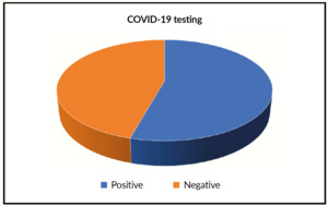
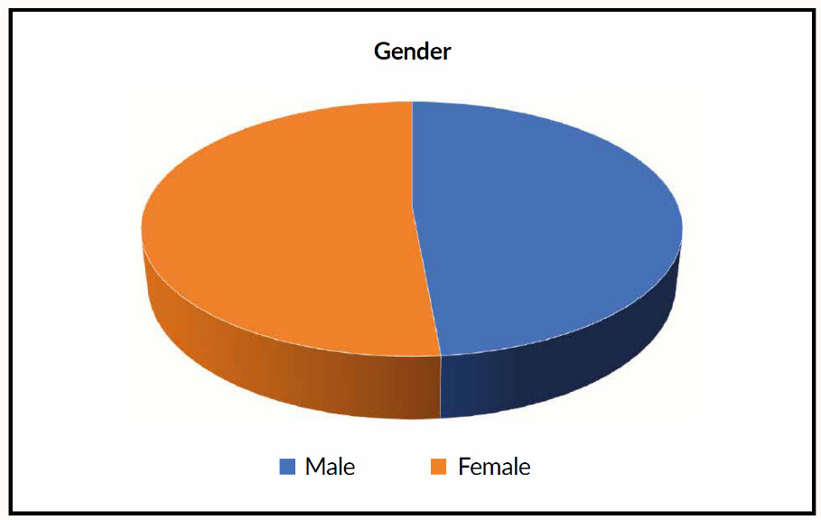
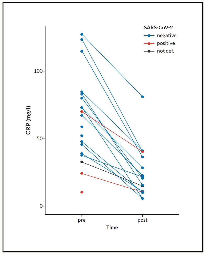
Comments: There is a significant decrease of CRP in all patients receiving Nigella sativa (NS). Both test t-test and Wilcoxon signed-rank test gives p-values <0.001. We included 35 patients (male 45.5%, female 54.5) (Figure 2). Age (mean+/-SD) was 51.79+/-17.32 years. CRP fall from 63.48+/-32.94 mg/L to 32.94+/-23.10 mg/L. SARS positive cases were 19 (55.9%) of whole tested patients (Figure 3). All patients had a significant reduction of their respective symptoms.

Figure 2
Figure 2.Number of people testing positive by gender.
Figure 3
Figure 3.Number of people who tested positive for COVID-19.
Figure 4
Figure 4.Effect of NS Treatment on CRP.

CRP, C-reactive protein; NS, Nigella sativa.

Discussion

Most of the medicinal properties of N. sativa were explored previously as an orally provided nutritional or dietary supplement of ground seeds in tablet or capsule form, oil extract in soft gel capsules, or as an aqueous extract in the form of fluid.24 Different combinations of N. sativa powder or extract with other vitamins such as vitamin E, are commonly offered as black seeds extract or black cumin oil to enhance natural physiological and immunological defense.

Extracts of N. sativa and specifically TQ, exhibited a broad antimicrobial range, including bacteria, viruses, and fungi. However, their effectiveness is dependent on the species of the target microorganisms.25 The mechanism of the antimicrobial effect of N. sativa has not been reported until now. Its antimicrobial property could be attributed to the active constituents particularly TQ and melanin.26 Their broad spectrum of activity may be the reason that the key processes of the organisms are affected.27

Endothelins are potent regulators of vascular tone, which also have mitogenic, apoptotic, and immunomodulatory properties.28,29 Three isoforms of endothelin have been identified to date, with endothelin-1 (ET-1) being considered the best route of study. ET-1 is classically considered a potent vasoconstrictor. However, in addition to the effects of ET-1 on vascular smooth muscle cells, the peptide is increasingly recognized as a proinflammatory cytokine.29,30 ET-1 causes platelet aggregation and plays a role in the increased expression of leukocyte adhesion molecules, the synthesis of inflammatory mediators contributing to vascular dysfunction. High levels of ET-1 are found in alveolar macrophages, leukocytes, and fibroblasts.30,31 Clinical and experimental data indicate that ET-1 is involved in the pathogenesis of sepsis,32,33 viral and bacterial pneumonia.34,35 Rickettsia conorii infections,36 Chagas disease,37,38 and severe malaria.39,40 A post-mortem histological analysis study of COVID-19 patients in Zurich revealed viral inclusion structures in endothelial cells. This histological study showed endotheliitis in many organs of the above-mentioned patients. In addition to that, endothelial dysfunction is a principal determinant of microvascular dysfunction by shifting the vascular equilibrium towards more vasoconstriction with subsequent organ ischemia, inflammation with associated tissue edema, and procoagulant state.41 A study has explained by their findings that the presence of viral elements within endothelial cells and an accumulation of inflammatory cells, with evidence of endothelial and inflammatory cell death. These findings suggest that SARS-CoV-2 infection facilitates the induction of endotheliitis in several organs as a direct consequence of viral involvement and of the host inflammatory response (Figure 5). In addition, induction of apoptosis and pyroptosis might have an important role in endothelial cell injury in patients with COVID-19.42 N. sativa has a reducing effect on oxidative like endothelin and it reduces the endothelin level in bronchoalveolar lavage and lung tissue.43

Figure 5
Figure 5.Endotheliitis. Adapted from Varga et al. 2020.42

A, B) Electron microscopy of kidney tissue shows viral inclusion bodies in a peritubular space and viral particles in endothelial cells of the glomerular capillary loops. Aggregates of viral particles (arrow) appear with dense circular surface and lucid center. The asterisk in panel B marks peritubular space consistent with capillary containing viral particles. The inset in panel B shows the glomerular basement membrane with endothelial cell and a viral particle (arrow; about 150 nm in diameter). C) Small bowel resection specimen of patient 3, stained with hematoxylin and eosin. Arrows point to dominant mononuclear cell infiltrates within the intima along the lumen of many vessels. The inset of panel C shows immunohistochemical staining of caspase 3 in small bowel specimens from a serial section of tissue described in panel D.Staining patterns were consistent with apoptosis of endothelial cells and mononuclear cells observed in the hematoxylin-eosin stained sections, indicating that apoptosis is induced in a substantial proportion of these cells. D) Post-mortem lung specimen stained with hematoxylin and eosin showed thickened lung septa, including a large arterial vessel with mononuclear and neutrophilicin filtration (arrow in upper inset). The lower inset shows immunohistochemical staining of caspase 3 on the same lung specimen; these staining patterns were consistent with apoptosis of endothelial cells and mononuclear cells observed in the hematoxylin-eosin-stained sections.

ACE2 has been identified as the functional receptor for SARS‐CoV.44 Li et al. showed that ACE2 can be immunoprecipitated by the S1 domain of the SARS‐CoV virus and that ACE2 can promote viral replication.

The antihypertensive effect of N. sativa is mediated by a reduction in cardiac oxidative stress and angiotensin-converting enzyme activity, an increase in cardiac heme oxygenase-1 activity, and prevention of plasma nitric oxide loss. Thus, N. sativa might be beneficial for controlling hypertension.45 It has an inhibitory effect of angiotensin-converting enzyme (ACE) that inhibits not only the conversion of angiotensin II from angiotensin I but also the bradykinase that degrades bradykinin (Figure 6). Bradykinin is generally a proinflammatory or an inflammatory mediator, which may inhibit viral replication.

SARS-CoV-2 has been shown to bind to angiotensin-converting enzyme 2 (ACE 2) to enter human cells by binding via the S protein on its surface. During infection, the S protein is cleaved into subunits S1 and S2. The S1 contains the receptor-binding domain (RBD), which allows coronaviruses to directly bind to the peptidase domain (PD) of ACE 2, and the S2 is believed to have a significant role in membrane fusion.46 Through the understanding of the ACE 2, it is expected to lead to producing antivirus or a vaccine that can block coronavirus infection by targeting ACE 2.

Figure 6
Figure 6.Possible mechanisms for the actions of NS against COVID-19.

NS, Nigella sativa; COVID-19, coronavirus disease 2019.

Take-Home Messages

  • Currently, there is a quite number of treatment options for viral infections with SARS-CoV-2 available. The majority of treatment options are of a supportive nature, and none is an absolute satisfactory therapeutic management. Thus, there are challenging further trends and trials for viral infection treatment management with SARS-CoV-2, with minimal or no side effects.

  • N. sativa exhibits the potential to be a distinguished drug for treating COVID-19 and different microbial infections. A pilot clinical study using N. sativa with big data and analytics is highly in demand based on the data presented above, which will propagate its unique properties to COVID-19 treatment management.


Disclosure statement

The authors have not declared any financial or personal connections in connection with this post.

Acknowledgments

The authors would like to thank Fabio Valeri for assistance with statistical analysis and careful review of the case report.

Additional Information

https://cdn.healthbook.network/wp-content/uploads/2021/02/210201-healthbook-TIMES-Fetian-IR-et-al_appendix.pdf

Accepted: March 02, 2021 CEST

References

1.
Chen N, Zhou M, Dong X, et al. Epidemiological and clinical characteristics of 99 cases of 2019 novel coronavirus pneumonia in Wuhan, China: a descriptive study. Lancet. 2020;395(10223):507-513. doi:10.1016/S0140-6736(20)30211-7
Google Scholar
2.
Wang D, Hu B, Hu C, et al. Clinical Characteristics of 138 Hospitalized Patients With 2019 Novel Coronavirus-Infected Pneumonia in Wuhan, China. JAMA. 2020;323(11):1061-1069. doi:10.1001/jama.2020.1585
Google Scholar
3.
Li Q, Guan X, Wu P, et al. Early Transmission Dynamics in Wuhan, China, of Novel Coronavirus-Infected Pneumonia. N Engl J Med. 2020;382(13):1199-1207. doi:10.1056/NEJMoa2001316
Google Scholar
4.
World Health Organization. COVID 19 Public Health Emergency of International Concern (PHEIC). Global research and innovation forum: towards a research roadmap. 2020. Accessed January 2021. https:/​/​covid19-evidence.paho.org/​handle/​20.500.12663/​714
5.
Covid-19 Coronavirus Pandemic. Worldometer; 2021. Accessed January 2021. https:/​/​www.worldometers.info/​coronavirus/​?%3D%3D
6.
Cyranoski D. ‘We need to be alert’: Scientists fear second coronavirus wave as China’s lockdowns ease. Nature. Published online March 30, 2020:d41586-020-00938-0.
7.
Singhal T. A Review of Coronavirus Disease-2019 (COVID-19). Indian J Pediatr. 2020;87(4):281-286. doi:10.1007/s12098-020-03263-6
Google Scholar
8.
Pakdemirli E, Mandalia U, Monib S. Positive Chest CT Features in Patients With COVID-19 Pneumonia and Negative Real-Time Polymerase Chain Reaction Test. Cureus. 2020;12(8):e9942. doi:10.7759/cureus.9942
Google Scholar
9.
Ferran M. Coronavirus tests are pretty accurate, but far from perfect. The Conversation. Accessed January 2021. https:/​/​theconversation.com/​coronavirus-tests-are-pretty-accurate-but-far-from-perfect-136671
10.
Tan C, Huang Y, Shi F, et al. C‐reactive protein correlates with computed tomographic findings and predicts severe COVID‐19 early. J Med Virol. 2020;92(7):856-862. doi:10.1002/jmv.25871
Google Scholar
11.
Tian W, Jiang W, Yao J, et al. Predictors of mortality in hospitalized COVID-19 patients: A systematic review and meta-analysis. J Med Virol. 2020;92(10):1875-1883. doi:10.1002/jmv.26050
Google Scholar
12.
Wang G, Wu C, Zhang Q, et al. C-Reactive Protein Level May Predict the Risk of COVID-19 Aggravation. Open Forum Infect Dis. 2020;7(5):ofaa153. doi:10.1093/ofid/ofaa153
Google Scholar
13.
Ali N. Is SARS-CoV-2 associated with liver dysfunction in COVID-19 patients? Clin Res Hepatol Gastroenterol. 2020;44(4):e84-e86. doi:10.1016/j.clinre.2020.05.002
Google Scholar
14.
Lagunas-Rangel FA. Neutrophil-to-lymphocyte ratio and lymphocyte-to-C-reactive protein ratio in patients with severe coronavirus disease 2019 (COVID-19): A meta-analysis. J Med Virol. 2020;92(10):1733-1734. doi:10.1002/jmv.25819
Google Scholar
15.
Marnell L, Mold C, Du Clos TW. C-reactive protein: ligands, receptors and role in inflammation. Clin Immunol Orlando Fla. 2005;117(2):104-111. doi:10.1016/j.clim.2005.08.004
Google Scholar
16.
Young B, Gleeson M, Cripps AW. C-reactive protein: a critical review. Pathology (Phila). 1991;23(2):118-124. doi:10.3109/00313029109060809
Google Scholar
17.
Pepys MB, Hirschfield GM. C-reactive protein: a critical update. J Clin Invest. 2003;111(12):1805-1812. doi:10.1172/JCI18921
Google Scholar
18.
Ballou SP, Kushner I. C-reactive protein and the acute phase response. Adv Intern Med. 1992;37:313-336.
Google Scholar
19.
Chen C, Zhang Y, Huang J, et al. Favipiravir versus Arbidol for COVID-19: A Randomized Clinical Trial. Infectious Diseases (except HIV/AIDS). Published online March 20, 2020. doi:10.1101/2020.03.17.20037432
Google Scholar
20.
Chen, et al. Thalidomide Combined with Low-dose Glucocorticoid in the Treatment of COVID-19 Pneumonia. Preprints. Published online 2020:2020020395.
Google Scholar
21.
Fetian, et al. Nigella sativa Immunomodulatory Activity as a Potential Treatment of Novel Coronavirus (2019-nCoV) – A Review of Current Literature. healthbook Times. Accessed January 2021. http:/​/​cdn.healthbook.network/​pim/​2020/​10/​5e070689-7b42-4638-acae-16d1b71eeb8c.pdf
Google Scholar
22.
Tavakoly R, Arab A, Vallianou N, et al. The effect of Nigella sativa L. supplementation on serum C-reactive protein: A systematic review and meta-analysis of randomized controlled trials. Complement Ther Med. 2019;45:149-155. doi:10.1016/j.ctim.2019.06.008
Google Scholar
23.
Bari SF, Khan A, Lawson T. C reactive protein may not be reliable as a marker of severe bacterial infection in patients receiving tocilizumab. BMJ Case Rep. 2013;2013. doi:10.1136/bcr-2013-010423
Google Scholar
24.
Dajani EZ, Shahwan TG, Dajani NE. Overview of the preclinical pharmacological properties of Nigella sativa (black seeds): a complementary drug with historical and clinical significance. J Physiol Pharmacol Off J Pol Physiol Soc. 2016;67(6):801-817.
Google Scholar
25.
Forouzanfar F, Bazzaz BSF, Hosseinzadeh H. Black cumin (Nigella sativa) and its constituent (thymoquinone): a review on antimicrobial effects. Iran J Basic Med Sci. 2014;17(12):929-938.
Google Scholar
26.
Bakathir HA et al. Detection of the antibacterial effect of nigella sativa ground seedswith water. Afr J Tradit Complement Altern Med. 2011;8(2):159-164.
Google Scholar
27.
Monika T et al. An investigational study of antibacterial activities of Nigella sativa on Mastits in dairy crossbred cows. Int J Adv Scient Tech Res. 2013;3(4):273-283.
Google Scholar
28.
Kedzierski RM, Yanagisawa M. Endothelin system: the double-edged sword in health and disease. Annu Rev Pharmacol Toxicol. 2001;41:851-876. doi:10.1146/annurev.pharmtox.41.1.851
Google Scholar
29.
Bagnato A, Loizidou M, Pflug BR, Curwen J, Growcott J. Role of the endothelin axis and its antagonists in the treatment of cancer. Br J Pharmacol. 2011;163(2):220-233. doi:10.1111/j.1476-5381.2011.01217.x
Google Scholar
30.
Sessa WC, Kaw S, Hecker M, Vane JR. The biosynthesis of endothelin-1 by human polymorphonuclear leukocytes. Biochem Biophys Res Commun. 1991;174(2):613-618. doi:10.1016/0006-291x(91)91461-k
Google Scholar
31.
Gu J, Pinheiro JM, Yu CZ, D’Andrea M, Muralidharan S, Malik A. Detection of endothelin-like immunoreactivity in epithelium and fibroblasts of the human umbilical cord. Tissue Cell. 1991;23(4):437-444. doi:10.1016/0040-8166(91)90002-b
Google Scholar
32.
Tschaikowsky K, Sägner S, Lehnert N, Kaul M, Ritter J. Endothelin in septic patients: effects on cardiovascular and renal function and its relationship to proinflammatory cytokines. Crit Care Med. 2000;28(6):1854-1860. doi:10.1097/00003246-200006000-00028
Google Scholar
33.
Goto T, Hussein MH, Kato S, et al. Endothelin receptor antagonist attenuates oxidative stress in a neonatal sepsis piglet model. Pediatr Res. 2012;72(6):600-605. doi:10.1038/pr.2012.134
Google Scholar
34.
Schuetz P, Stolz D, Mueller B, et al. Endothelin-1 precursor peptides correlate with severity of disease and outcome in patients with community acquired pneumonia. BMC Infect Dis. 2008;8:22. doi:10.1186/1471-2334-8-22
Google Scholar
35.
Samransamruajkit R, Moonviriyakit K, Vanapongtipagorn P, Prapphal N, Deerojanawong J, Poovorawan Y. Plasma endothelin-1 in infants and young children with acute bronchiolitis and viral pneumonia. Asian Pac J Allergy Immunol. 2002;20(4):229-234.
Google Scholar
36.
Davì G, Giammarresi C, Vigneri S, et al. Demonstration of Rickettsia Conorii-induced coagulative and platelet activation in vivo in patients with Mediterranean spotted fever. Thromb Haemost. 1995;74(2):631-634.
Google Scholar
37.
Petkova SB, Huang H, Factor SM, et al. The role of endothelin in the pathogenesis of Chagas’ disease. Int J Parasitol. 2001;31(5-6):499-511. doi:10.1016/s0020-7519(01)00168-0
Google Scholar
38.
Petkova SB, Tanowitz HB, Magazine HI, et al. Myocardial expression of endothelin-1 in murine Trypanosoma cruzi infection. Cardiovasc Pathol Off J Soc Cardiovasc Pathol. 2000;9(5):257-265. doi:10.1016/s1054-8807(00)00045-4
Google Scholar
39.
Dai M, Freeman B, Bruno FP, et al. The novel ETA receptor antagonist HJP-272 prevents cerebral microvascular hemorrhage in cerebral malaria and synergistically improves survival in combination with an artemisinin derivative. Life Sci. 2012;91(13-14):687-692. doi:10.1016/j.lfs.2012.07.006
Google Scholar
40.
Dietmann A, Lackner P, Helbok R, et al. Opposed circulating plasma levels of endothelin-1 and C-type natriuretic peptide in children with Plasmodium falciparum malaria. Malar J. 2008;7:253. doi:10.1186/1475-2875-7-253
Google Scholar
41.
Bonetti PO, Lerman LO, Lerman A. Endothelial dysfunction: a marker of atherosclerotic risk. Arterioscler Thromb Vasc Biol. 2003;23(2):168-175. doi:10.1161/01.atv.0000051384.43104.fc
Google Scholar
42.
Varga Z, Flammer AJ, Steiger P, et al. Endothelial cell infection and endotheliitis in COVID-19. Lancet. 2020;395(10234):1417-1418. doi:10.1016/S0140-6736(20)30937-5
Google Scholar
43.
Mazouchian H et al. The effect of thymoquinone, the main constituent of Nigella sativa, on endothelin level of ovalbumin sensitized guinea pigs. Adv Biores. 2013;4(5):105-108.
Google Scholar
44.
Li W, Moore MJ, Vasilieva N, et al. Angiotensin-converting enzyme 2 is a functional receptor for the SARS coronavirus. Nature. 2003;426(6965):450-454. doi:10.1038/nature02145
Google Scholar
45.
Jaarin K, Foong WD, Yeoh MH, et al. Mechanisms of the antihypertensive effects of Nigella sativa oil in L-NAME-induced hypertensive rats. Clin Sao Paulo Braz. 2015;70(11):751-757. doi:10.6061/clinics/2015(11)07
Google Scholar
46.
Tai W, He L, Zhang X, et al. Characterization of the receptor-binding domain (RBD) of 2019 novel coronavirus: implication for development of RBD protein as a viral attachment inhibitor and vaccine. Cell Mol Immunol. 2020;17(6):613-620. doi:10.1038/s41423-020-0400-4
Google Scholar

Appendix I

Appendix II

Summary of complete dataset

sex2 age crp1 crp2 sars2
female:18 Min. :16.00 Min. : 10.00 Min. : 5.2 negative:15
male :15 1st Qu.:39.00 1st Qu.: 38.25 1st Qu.:10.0 positive:19
NA’s : 2 Median :51.00 Median : 58.50 Median :15.3 NA’s : 1
Mean :51.79 Mean : 63.48 Mean :23.1
3rd Qu.:62.00 3rd Qu.: 81.35 3rd Qu.:36.2
Max. :77.00 Max. :127.20 Max. :81.2
NA’s :2 NA’s :16 NA’s :18
thday
Min. :2.000
1st Qu.:2.000
Median :3.000
Mean :2.765
3rd Qu.:3.000
Max. :4.000
NA’s :18

Summary of splitted dataset: SARS-CoV-2 negative/positive

a001a1_1$sars2: negative
sex2 age crp1 crp2 sars2
female:6 Min. :16.00 Min. : 37.5 Min. : 5.20 negative:15
male :9 1st Qu.:32.00 1st Qu.: 46.6 1st Qu.:10.00 positive: 0
Median :41.00 Median : 66.9 Median :20.20
Mean :43.13 Mean : 71.3 Mean :24.47
3rd Qu.:50.50 3rd Qu.: 83.6 3rd Qu.:36.20
Max. :77.00 Max. :127.2 Max. :81.20
NA’s :2
thday
Min. :2.000
1st Qu.:2.000
Median :3.000
Mean :2.833
3rd Qu.:3.000
Max. :4.000
NA’s :3

a001a1_1$sars2: positive
sex2 age crp1 crp2 sars2
female:11 Min. :31.00 Min. :10.00 Min. :10 negative: 0
male : 6 1st Qu.:50.00 1st Qu.:17.00 1st Qu.:10 positive:19
NA’s : 2 Median :59.00 Median :24.00 Median :10
Mean :58.24 Mean :34.67 Mean :20
3rd Qu.:72.00 3rd Qu.:47.00 3rd Qu.:25
Max. :77.00 Max. :70.00 Max. :40
NA’s :2 NA’s :16 NA’s :16
thday
Min. :2.0
1st Qu.:2.0
Median :2.5
Mean :2.5
3rd Qu.:3.0
Max. :3.0
NA’s :15

Statistics of the complete dataset
Overall
N 35
sex2 = male (%) 15 (45.5)
age (mean (SD)) 51.79 (17.32)
crp1 (mean (SD)) 63.48 (32.94)
crp2 (mean (SD)) 23.10 (19.43)
sars2 = positive (%) 19 (55.9)
Statistics of splitted dataset: SARS-CoV-2 negative/positive
negative positive p test
N 15 19
sex2 = male (%) 9 (60.0) 6 (35.3) 0.297
age (mean (SD)) 43.13 (17.43) 58.24 (13.94) 0.011
crp1 (mean (SD)) 71.30 (30.70) 34.67 (31.39) 0.078
crp2 (mean (SD)) 24.47 (21.05) 20.00 (17.32) 0.739
sars2 = positive (%) 0 (0.0) 19 (100.0) <0.001