Introduction
Primary lymphedema is a rare genetic disorder with a prevalence of approximately 1 in 10,000, that affects the development and function of the lymphatic system. It can occur as an isolated defect or as part of a genetic syndrome, and is classified into three types based on the age of onset: congenital lymphedema, lymphedema praecox, and lymphedema tarda.1,2 Recent studies have investigated the genetic and molecular basis of primary lymphedema, and have identified several genes and signaling pathways involved in lymphatic development and function. For example, mutations in the VEGFR3 gene and other genes have been linked to primary lymphedema.1,3,4 In addition to genetic factors, other risk factors for primary lymphedema include obesity, trauma, infection, and cancer. Treatment options for primary lymphedema include compression therapy, manual lymphatic drainage, and surgery, but there is still a need for more effective and personalized therapies.1
The current treatment options for primary lymphedema mainly focus on reducing symptoms and improving patients’ quality of life. Conservative management includes compression therapy, which involves the use of bandages, stockings or sleeves to apply pressure to the affected limb and reduce swelling. Manual lymphatic drainage, a specialized massage technique to stimulate the lymphatic system, is also used in combination with compression therapy. The combination technique is termed complex decongestive therapy (CDT).1 However, these treatments have limitations in terms of efficacy and long-term compliance. Surgical options include lymphaticovenular anastomosis (LVA), lymph node transfer (LNT) and debulking procedures, which aim to redirect or bypass lymphatic flow and remove excess tissue.5 These surgical options have shown promising results in reducing lymphedema severity and improving patient outcomes, although they are associated with surgical risks and complications. The prognosis for primary lymphedema varies depending on the type and severity of the condition, as well as the patient’s age and overall health. Long-term management and regular follow-up are recommended to prevent disease progression and manage complications. Research into new treatments and personalized therapies for primary lymphedema is ongoing. Overall, the management of primary lymphedema remains a challenge, and a multidisciplinary approach is required to optimize patient care.1,3
Correlation with retinal Edema and Rescue: Hypothesis and Rational for Treatment Choice
Retinal edema, as it presents in cases of normal tension glaucoma, can be driven by an increase in retinal venous pressure (RVP).6 RVP is actively regulated by endothelin and other vasoconstrictors, with increased endothelin causing an increase in RVP that can lead to edema. Endothelin, which can originate from local tissues, increases intraluminal pressure by increasing outflow resistance, contracting retinal veins in particular where they exit the eye (for review of this mechanism in retina, see also https://glaucomaresearch.ch/en/retinal-venous-pressure).7,8
Elevated RVP has been associated with endothelial dysfunction and impaired vascular regulation, with recent studies indicating that diminished patency of blood vessels plays a key role in the increase of retinal venous pressure and subsequent edema.8 Nutritional supplementation with several B vitamins has been shown to improve endothelial function and reduce symptoms in patients with elevated RVP.9,10 Specifically, a combination of B vitamins and minerals has been found to enhance blood vessel dilation, reduce oxidative stress, and improve blood flow regulation. These findings suggest that endothelial dysfunction may be reversible with appropriate nutritional intervention.
Since endothelin similarly contracts lymphatic vessels, we speculate that a similar mechanism may be at play in the pathogenesis of primary lymphedema.11 Specifically, we propose that endothelin may constrict lymphatic veins, particularly in the prenodal regions, with the resultant weakening of the lymphatic vessel walls and elevated intraluminal pressure reducing lymphatic drainage and contributing to edema due to the accumulation of lymphatic fluid. If this hypothesis is correct, it may be possible to improve lymphatic vessel function with nutritional supplementation. Specifically, we suggest that a combination of B vitamins and minerals may be effective in enhancing lymphatic vessel patency and reducing the severity of primary lymphedema symptoms.12 This approach could potentially reduce the need for compression stockings, which are currently a mainstay of primary lymphedema treatment. Further research is needed to investigate the potential of nutritional supplementation for improving lymphatic vessel function in primary lymphedema patients.
In addition to the nutritional interventions described above, Flammer and colleagues have also proposed the use of plasma homocysteine levels as a biomarker for endothelial health.9,13 Homocysteine is a non-protein amino acid that has been implicated in the development of endothelial dysfunction and cardiovascular disease. Plasma homocysteine levels have been shown to be significantly elevated in patients with normal tension glaucoma and serve as a biomarker for disease severity.10 Specifically, plasma homocysteine levels over 10 µmol/L are associated with deteriorating vascular health, while levels of 10 µmol/L and under are associated with good vascular health. These findings suggest that homocysteine levels may be a useful tool for identifying patients at risk of endothelial dysfunction and guiding nutritional interventions.
This approach may have potential in the context of primary lymphedema. As noted above, recent research has documented that elevated plasma homocysteine levels serve as a biomarker for vascular disease severity.10 If confirmed in lymphatic vessels, this finding could have important implications for the diagnosis and management of primary lymphedema, and provide a valuable tool for monitoring disease progression and response to treatment. Further research is needed to investigate the potential of plasma homocysteine levels as a biomarker for primary lymphedema. Recent evidence links elevated homocysteine levels with decreased vascular health or increased blood pressure.9,14 We postulated that this may be true for homocysteine and lymphatic health and pressure.
Given our experience with using a high dose medicinal food (food for special medical purposes, FSMP) in treatment of retinal vascular conditions such as glaucoma and macular degeneration, we chose to use the same product developed for retinal vascular conditions in the case study reported here.15 Ocufolin is a food for special medical purposes (FSMP) containing vitamins B1 (1.5 mg), B2 (10 mg), B6 (3 mg), B12 (500 μg), L-methyl folate calcium (900 μg), as well as several minerals and AREDS2 vitamins. The B vitamins listed have been shown to be the ones necessary for restoring endothelial cell health to ensure adequate microvascular flow (Flammer, 2021), and we chose to use Ocufolin for this case to provide a single supplement that could confirm our hypothesis that edema in primary lymphedema may be reduced in a similar manner to that used in retina. Additionally, a dietary source of omega-3 fatty acids was included biweekly. A primary lymphedema patient using this FSMP may be expected to have a similar improvement in lymphatic venous drainage and concomitant reduction in edema as that seen when using Ocufolin in treatment of retinal vascular disease.9,15
Patient Information
The case involved a 62-year-old male with primary lymphedema, with age of onset at 53. The primary lymphedema diagnosis was made by physicians at Massachusetts General Hospital, following immunological, vascular, radiological, and infectious disease testing. The Lymphedema Center of Beth Israel Hospital (Boston) confirmed diagnosis and began treatment of compression garments and manual lymph drainage (multiple times), advancing to compressive wrapping.
Subject volunteered to take a high-dose multivitamin for a year. The patient had been experiencing primary lymphedema in both legs, requiring full-length (thigh-high) compression stockings every day, with knee high compression stockings every night. The patient reported his mother suffered from a similar condition, and neither his nor his mother’s resolved with the compression garments or modifications to diet or use of diuretics. The compression garments were to contain the evident edema and had no effect in reducing the edema.
Intervention & Assessment
The patient began taking a high-dose multivitamin, Ocufolin, which is designated a “food for special medical purposes” (FSMP) in Europe, and termed “medicinal food” in the US, due to its high levels of B vitamins. Ocufolin is produced in Switzerland by Aprofol and in the US by GHF and was purchased from GHF for use. The patient took the dose listed on the packaging for 12 months, and included fatty fish biweekly in his diet as the source of omega 3 fatty acids. He did not have any oral omega-3 substitution. Prior to taking the vitamin, the patient had plasma homocysteine levels of 11.8 µmol/ml, which indicated deteriorating vascular health. After six months of taking Ocufolin, the patient’s homocysteine level was 11.0 µmol/ml. However, at 12 months the patient’s homocysteine levels had decreased to 9.6 µmol/ml, which correlates with good vascular health. While this is only a single case and further research is recommended to confirm these results, it does suggest that nutritional interventions may hold promise for the management of primary lymphedema.
In addition to the improvement in homocysteine levels (Table 1), the patient in the case study also experienced a persistent reduction in swelling in both legs while taking Ocufolin. Average measurements of right and left thighs decreased from 59 cm to 53 cm after 12 months, while average measurements of right and left calves decreased from 47 cm to 41 cm (Table; daily variation continues to be approximately 1 cm). Three months into taking the vitamin, the patient had diminished swelling in both legs and was able to ambulate without full-length compression stockings for an afternoon without complication. After six months on daily Ocufolin, the patient had further reduction in swelling of both legs and was able to go without compression stockings for a full day of moderate activity. There was a concomitant decrease in body weight of approximately 5 kg from start of trial to completion, presumed to be a function of the decreased edema.
The patient has continued taking Ocufolin. At 12 months he reported no longer wearing full length compression stockings at all, using knee high compression stockings most days (average of 5 days/wk) or diabetic socks (average of 2 days/wk), with many days using no socks until mid-afternoon. He engaged in light hiking and swimming activities without need for any compression, and found that he only needed compression stockings when sitting for a prolonged period (over 30 min), as he found no swelling increase when active. He was also able to sleep at night without any compression, with no adverse effects.
At 28 months, the patient continues taking Ocufolin. He reports no longer using the full length (thigh high) compression stockings, and has not used them at all for two years. He uses knee high compression stockings on occasion, estimating that he uses the light weight (30 denier) knee highs on alternate days or alternate nights, depending on activity. Specifically using them at night prophylactically in advance of a day of activity in shorts and sandals, when he does not wear any stockings during such days. He engages in outdoor recreation without restriction. He further reports that all his pants fit now, that he can walk with ease and stability with no ambulatory insecurity, that he wears normal shoes now, and has experienced no further episodes of slipping now that sensation from his feet is unimpeded by edema.
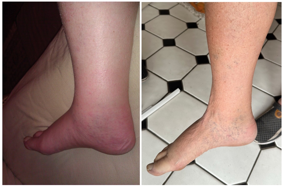
Figure 1 is a photo-montage of the patient’s right leg, with the left image taken at the beginning of the trial and the right image taken recently.

Throughout the course of this case study, the patient reported that there were no issues with the daily supplement, rather he stated that the joy of seeing improvement motivated him to take the daily Ocufolin and to continue it well past the study end date. Additionally, there are no reported tolerability concerns for Ocufolin in the literature, nor any reported adverse outcomes.
Discussion
While this case study is limited, it does suggest that nutritional supplementation with high levels of B vitamins may be a promising treatment for primary lymphedema. Further study is needed to confirm these findings and to explore the mechanisms by which nutritional interventions may improve lymphatic vessel function. It may be that such work can discern whether vitamin supplementation may benefit cases of secondary lymphedema, which merits similar study.
Because there are no adverse impacts from the high levels of B vitamin supplementation, this case offers hope that such therapy may be an effective first line treatment for primary lymphedema, perhaps as an adjunct to the current standard of compression garments and lymphatic massage. Should this be effective for others, adherence to the protocol will be easy, as each patient finding improvement in mobility will most likely choose to continue the vitamins.
Conclusion
Primary lymphedema can be successfully managed with high levels of certain B vitamins. These vitamins are necessary for restoring endothelial cell health in Flammer Syndrome, and appear to be having a similar effect for lymphatic vessels, restoring normal lymphatic drainage.
Ethics approval and consent to participate
General written consent was obtained from the patient for the publication of this study.
Availability of data and materials
All patient data supporting this article are included in anonymized form in the published article.
Conflicts of Interest
The author has declared that the study was conducted in the absence of any commercial or financial relationships that could be construed as a potential conflict of interest.
Funding
The author has declared that no financial support has been received from any organization for the submitted work.
Author Contribution
The authors wrote the manuscript and approved it.
Acknowledgements
The author thanks Prof Dr Josef Flammer for his review of the concept and recommendation to include omega-3 fatty acids in the diet.